-
Notifications
You must be signed in to change notification settings - Fork 16
Analyze Spheroid Cell Invasion In 3D Matrix
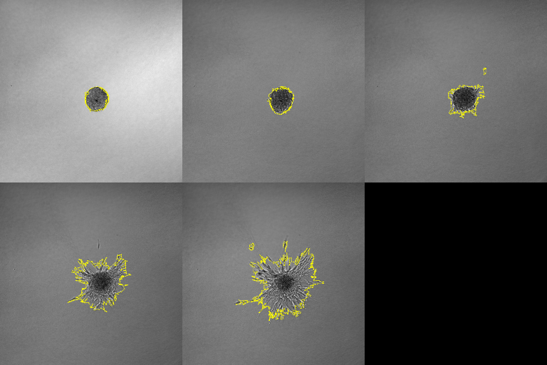
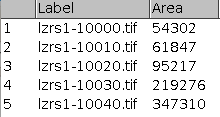
The tool allows to measure the area of the invading spheroïd in a 3D cell invasion assay. It can also count and measure the area of the nuclei within the speroïd. You can find an example time-series of image here: lzrs1.zip.
You can find the source code here.
The macro needs MorphoLibJ to be installed. In FIJI MorphoLibJ can be installed via the updater by activating the IJPB plugins site. To install the tools, drag the link: Analyze_Spheroid_Cell_Invasion_In_3D_Matrix.ijm to the ImageJ launcher window, save it under macros/toolsets in the FIJI installation and restart FIJI.
Select the Analyze_Spheroid_Cell_Invasion_In_3D_Matrix toolset from the >> button of the ImageJ launcher.
- the first button opens this help page
- the m button measures the area of the spheroid on the active image
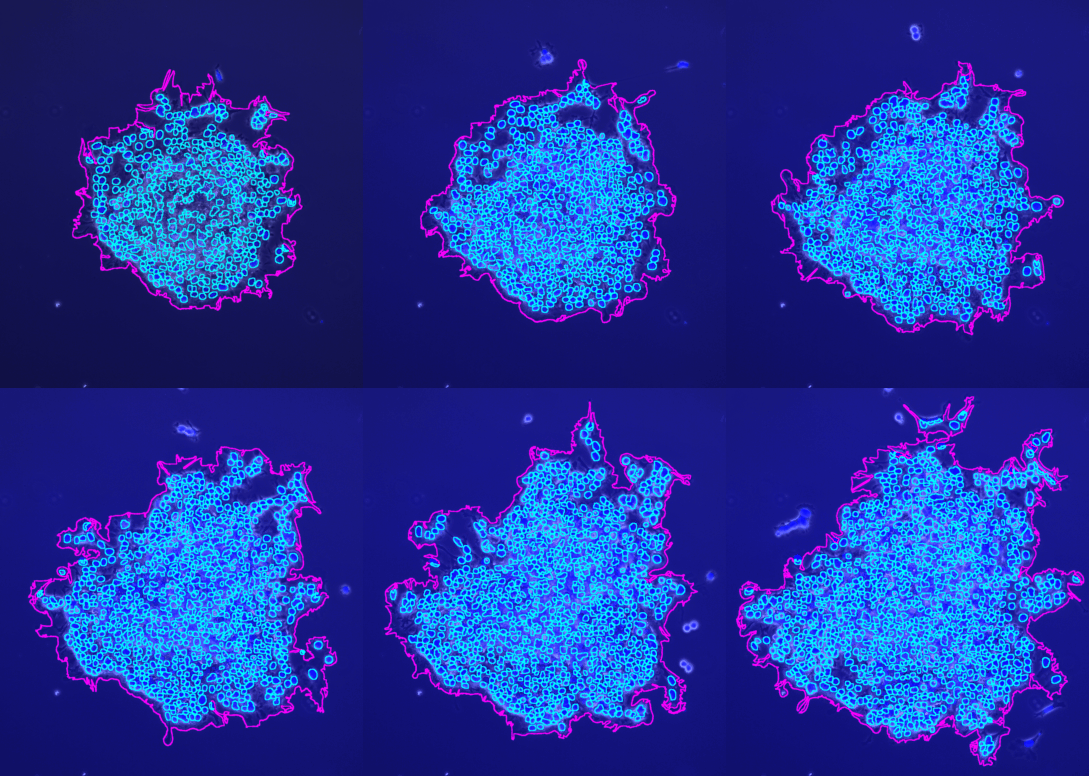
- the s button measures the area of the spheroid on a stack - it can also count the number of nuclei within the spheroid
- the b button runs the measurement of the spheroid in batch mode on a time-series of images
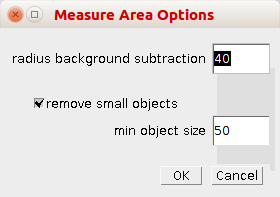
By right-clicking on the m button you can open the options dialog.
- radius background subtraction: the rolling ball radius for the subtract background command
- remove small objects: if selected small objects will be removed from the result
-
min. object size: the min. size of objects that will be taken into account when
remove small objectsis selected
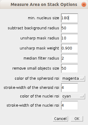
By right-clicking on the m button you can open the options dialog for processing a stack.
- min. nucleus size: minimum size of objects that are considered to be nuclei
- subtract background radius: the radius of the rolling ball for the subtract background operation
- unsharp mask radius: the radius of the unsharp mask
- unsharp mask weight: the weight of the unsharp mask
- median filter: the radius of the median filter
- remove small objects size: object smaller than this size are removed in the pre-processing
- color of the spheroid roi: the color of the roi that represents the outline of the spheroid
- stroke-width of the spheroid roi: the stroke-width of the roi, augment to get a thicker outline
- color of the nuclei roi: the color of the roi that represents the outlines of the nuclei
- stroke-width of the nuclei roi: the stroke-width of the roi, augment to get a thicker outline
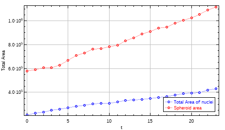
The s-button allows to analyse a film with 2 channels. The first channel must contain an image with a fluorescent staining of the nuclei and the second channel the spheroïd.
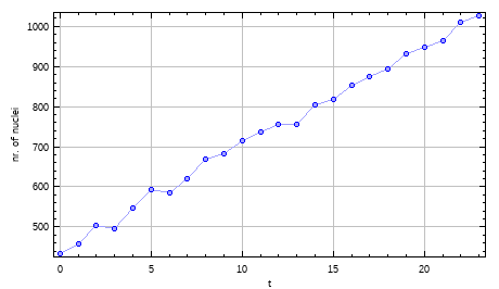
- Count: the number of detected nuclei in the frame (slice).
- Total Area: the total area of the detected nuclei in the frame (slice).
- Average Size: the average of the area of the nuclei in the frame (slice).
- spheroid area: the area of the spheroid in the frame (slice).
The image containing the rois as overlays and the results-table are saved into the results folder, the user specifies when running the tool.
-
Van Treuren, T., and Vishwanatha, J.K. (2018). CRISPR deletion of MIEN1 in breast cancer cells. PLoS ONE 13, e0204976.
-
Sperlich, J., and Teusch, N. (2018). Pseudopterosin Inhibits Proliferation and 3D Invasion in Triple-Negative Breast Cancer by Agonizing Glucocorticoid Receptor Alpha. Molecules 23, 1992.


 Volker Bäcker